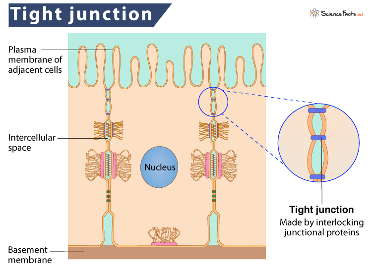
Epithelial cells line the outer surfaces of organs and blood vessels throughout the body and the inner cavities of various internal organs. So, tight junctions are also present in multiple locations. For example, skin, nephron, liver, bile duct, blood-brain-barrier, etc., to name a few.
Structure and Formation
Functions
A few transmembrane proteins (Claudins, Occludins, Junctional adhesion molecules, and Zonula occludens) from each cell membrane form these junctional complexes. They spread from the two adjacent membranes and interlock across the intercellular space. Along with these proteins, specific cytoplasmic adaptor proteins such as cingulin, PAR3, PAR6, and MUPP1 are also necessary for correctly organizing the integral membrane components of the junction. Though the main structural components of the junctions have been detected, it is still unclear how the formation is regulated. Upon complete formation and maturation, the junction appears like a series of local seals joined together in a maze-like fashion rather than a continuous seal. This barrier mechanism of tight junctions plays a regulatory role in controlling diseases like jaundice, edema, blood-borne metastasis, and diarrhea. 2. Maintaining cell polarity: They help maintain cell polarity by preventing the lateral diffusion of integral membrane proteins between the apical and basal surfaces. Comment * Name * Email * Website Save my name, email, and website in this browser for the next time I comment.
Δ
