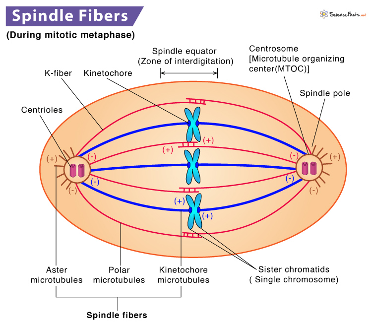
Spindle fibers play a significant role during nuclear division. They are responsible for the separation of sister chromatids during mitotic and meiotic cell division.
When Do They First Become Visible
Structure
Its Formations
Types of Mitotic Spindle
Functions
They are spindle-shaped fibers composed of 97% motor protein (tubulin) and 3% RNA. The microtubules are a polymer of 𝜶 and 𝞫-tubulin dimers. Spindle fibers, associated proteins, condensed chromosomes, and centrosomes or asters together form the spindle apparatus. In mitosis, these filaments form at opposite poles of the cell and meet at the equatorial plane. Together they produce a spindle-shaped structure, which is widest at the middle and tapered towards both ends. These are called spindle fibers.
Prophase
Spindle fibers start to form the centrioles at the opposite poles of the cell. In animal cells, the mitotic spindle appears as asters that surround each centriole pair. The sister chromatids attach to spindle fibers at their kinetochores during this phase. Also, the cell elongates as spindle fibers stretch from each pole.
Metaphase
As the mitotic spindle extends from each pole, they create tension. As a result, chromosomes line up at the metaphase plate. Microtubules from opposite spindle poles get attach to the two sister chromatids of each chromosome. In metaphase, the spindle has attached all the chromosomes and arranged them at the middle of the cell, making it ready for division.
Anaphase
As the spindle fibers attach to a protein complex called kinetochores, they either generate or transduce force to segregate and move sister chromatids to their opposite poles. Interpolar microtubules help in the lengthening and elongation of the cell to provide room for cell division.
Telophase
During this phase, the separated sister chromatids (now known as daughter chromosomes) reach the opposite poles and get enclosed within two new nuclei. The spindle fibers are found to disappear.
Cytokinesis
The cytoplasm divides, and two new daughter cells are formed with an equal number of chromosomes. Spindle fibers ensure this process.
Chromosome Movement
Cell movement occurs when microtubules of spindle fibers interact with motor proteins. Motor proteins like dyneins and kinesins move along microtubules causing the spindle fibers to contract (shorten) or relax (elongate). The disassembly and reassembly of microtubules produce the chromosomal movement required for cell division. Spindle fibers help in chromosomal movement during cell division by attaching to centromeres (the primary constriction site of a chromosome). Kinetochores produce fibers that attach sister chromatids to the spindle fibers. Both the kinetochore and polar microtubules work together on segregating chromosomes. Spindle fibers that do not bind chromosomes during cell division extend from one cell pole to the other. These fibers overlap and push cell poles away from one another resulting in cell elongation, preparing the cell for cytokinesis.
