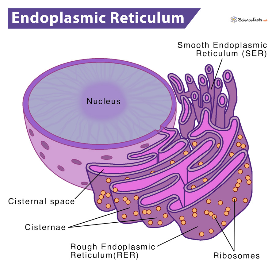
It is called ‘endoplasmic,’ as it is more concentrated in the inner side of the cytoplasm (endoplasm) than the outer side (ectoplasm). On the other hand, it is referred to as ‘reticulum’ due to its reticulate or network-like appearance. This kind of appearance can be observed under a light microscope. ER is absent in eggs, embryonic cells, mature RBC, and bacteria. Who Discovered Endoplasmic Reticulum In 1945, Porter and Thompson discovered Endoplasmic Reticulum. Later, in 1953, Keith Porter gave the name endoplasmic reticulum based on the observations made with the electron microscope on tissue culture cells.
Where Is it Located
Types of Endoplasmic Reticulum
Difference between Rough and Smooth Endoplasmic Reticulum
Structure and Characteristics
How are they Formed
Functions: What Does the Endoplasmic Reticulum Do
Rough endoplasmic reticulum (RER)Smooth endoplasmic reticulum (SER)
These two types of ER may be continuous with one another, plasma membrane and nuclear envelope.
Rough Endoplasmic Reticulum (RER)
These types of ER have a studded appearance due to the presence of ribosomes on their surface. For this reason, they are also called the granular endoplasmic reticulum. They have a series of connected flattened sacs having several ribosomes attached to their outer surface. They occur almost in all cells that are actively engaged in proteinsynthesis. For instance, they occur in liver cells, goblet cells, pancreatic cells, and plasma cells.
Smooth Endoplasmic Reticulum (SER)
Unlike RER, SER does not have any ribosomes attached to its outer surface, hence the name. It has a tubular form. It takes part in the production of the carbohydrate and chief lipids in cell membranes, phospholipids. It also commonly occurs in cells that synthesize steroid hormones (For example, Leydig cells in the testis and follicular cells in the ovary). It is also present in most neurons. SER transports the products of the RER to other cellular organelles and works together with the Golgi apparatus. ER consists of three components. They are cisternae, vesicles, and tubules.
- Lamellar Form (Cisternae):
Long, flattened, unbranched, sac-like structures arranged in parallel bundlesHave a diameter of 40-50 μmBear ribosomes on their surface, in the case of RERCommonly found in secretory cells
- Vesicular Form (Vesicle):
Ovoid or rounded structures and float freely in the cytoplasmHave a diameter of 25-500 μmAbundantly found in SEROccur at the end of cisternae and tubules
- Tubular Form (Tubules):
Smooth-walled and highly-branched tubular spaces, forming the reticular system along with the cisternae and vesiclesHave a diameter of 50-100 μmUsually occur in non-secretory cells like striated muscle cells
Composition: What is the Endoplasmic Reticulum Made out of
The membranes of ER contain high lipid content than its other protein partners. Some commonly found lipids are phospholipids, phosphatidylinositol, neutral lipids, sulfolipids, cholesterol, and some phytosterols. Nearly 30-40 different ER membrane proteins have been isolated. Out of which, many are enzymes like cytochrome P 450 and its subgroups, electron transport protein complexes like Cytochrome C reductase, Cytochrome b5 reductase. Glucose 6 phosphatases are also common in ER. Furthermore, though ER shows similarity in their structural appearances, they exhibit chemical heterogeneity at their cytoplasmic and lumen surfaces. They contain various resident proteins that perform various functions, like protein folding, protein modification, and protein transport. It has been observed that ER develops from plasma membranes, external nuclear membranes, and self-assembly. It is also true that both nuclear membrane and cell membrane are also derived from endomembranes. Also, all the other membrane-bound organelles have some connection with ER. All of them are involved in the synthesis of various liquid components and protein synthesis. Thus, they get self-assembled by incorporating required proteins and lipid and other fatty acid derivatives.
Protein Synthesis and Folding: Ribosomes, the protein factories attached to the ER, are more efficient in synthesizing protein than the free ribosomes lying in the cytoplasm. The ER then collects the process and transports the synthesized proteins to other parts of the cell.Lipid Formation: ER synthesizes triglycerides and phospholipids. It also stores lipids.Mechanical support: The ER divides the cell into different cytoplasmic compartments, providing mechanical support. Hence it is referred to as the cytoskeleton of the cell.Transport: ER acts as a circulatory system of a cell. It is involved in importing, exporting, and intracellular transportation of various substances, such as proteins, lipids, and enzymes.Synthesis of Cholesterol and Steroid Hormones: ER is the primary site for synthesizing cholesterol, the precursor for steroid hormones.Detoxification: It occurs in the ER of liver cells. It involves biochemical reactions by which harmful materials get converted into harmless substances suitable for excretion by the cell.Glycogenolysis: It is the process by which glycogen gets converted into glucose inside ER under the influence of an enzyme called glucose-6-phosphatase. This glucose gets transported to the blood.
