Giulio Bizzozero, an Italian pathologist, first discovered it. Josef Schaffer coined the term ‘desmosome’ in 1920.
Location of Desmosomes in a Cell
Structure
Functions of Desmosomes
Clinical Significances of Desmosomes
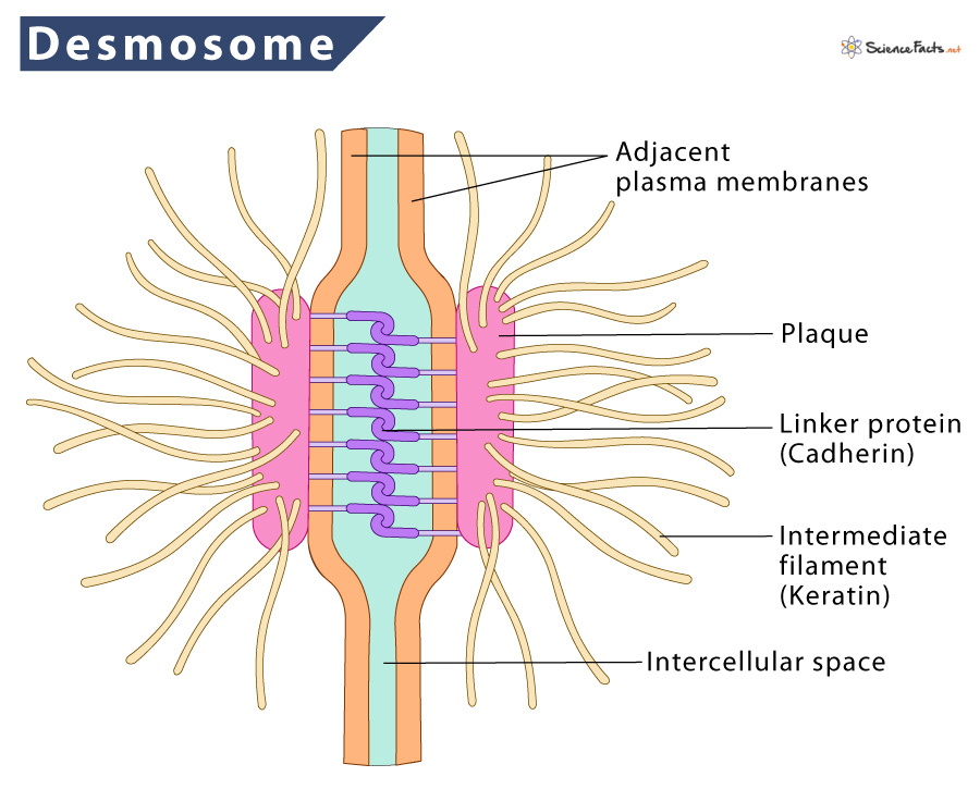
Desmosomes have a highly organized, electron-dense structure. They are less than one μm in diameter and consist of three parts.
What are Desmosomes Made of
They are composed of desmosome-intermediate filament complexes (DIFC), a network of cadherin proteins, linker proteins, and keratin intermediate filaments. A DIFC consists of three parts:
- Extracellular Core Region (Desmoglea) is approximately 34 nm in length containing the cadherin family of cell adhesion proteins – desmoglein and desmocollin.
- Outer Dense Plaque (ODP) is about 15–20 nm in length containing the intracellular ends of desmocollin and desmoglein, the N-terminus portion of desmoplakin, and the armadillo family proteins plakoglobin and plakophilin.
- Inner Dense Plaque (IDP) is about 15–20 nm in length containing the C-terminus portion of desmoplakin and their attachment to keratin intermediate filaments. Desmoplakin is most abundant in desmosome, acting as the moderator between the cadherin proteins in the plasma membrane and the keratin filaments Since desmosomes connect intermediate filaments of the cell cytoskeletons of adjacent cells, they provide mechanical strength to tissues. Also, they are found to affect the cadherin protein-based intracellular signaling pathways. Arrhythmogenic cardiomyopathy is usually caused due to mutations in desmoglein 2, but sometimes in desmocollin 2. Blistering disease such as pemphigus vulgaris (PV) is an autoimmune disease where auto-antibodies target desmogleins. PV is caused by circulating autoantibodies (IgG) that target Dsg3 (Desmoglein 3) and sometimes Dsg1.
