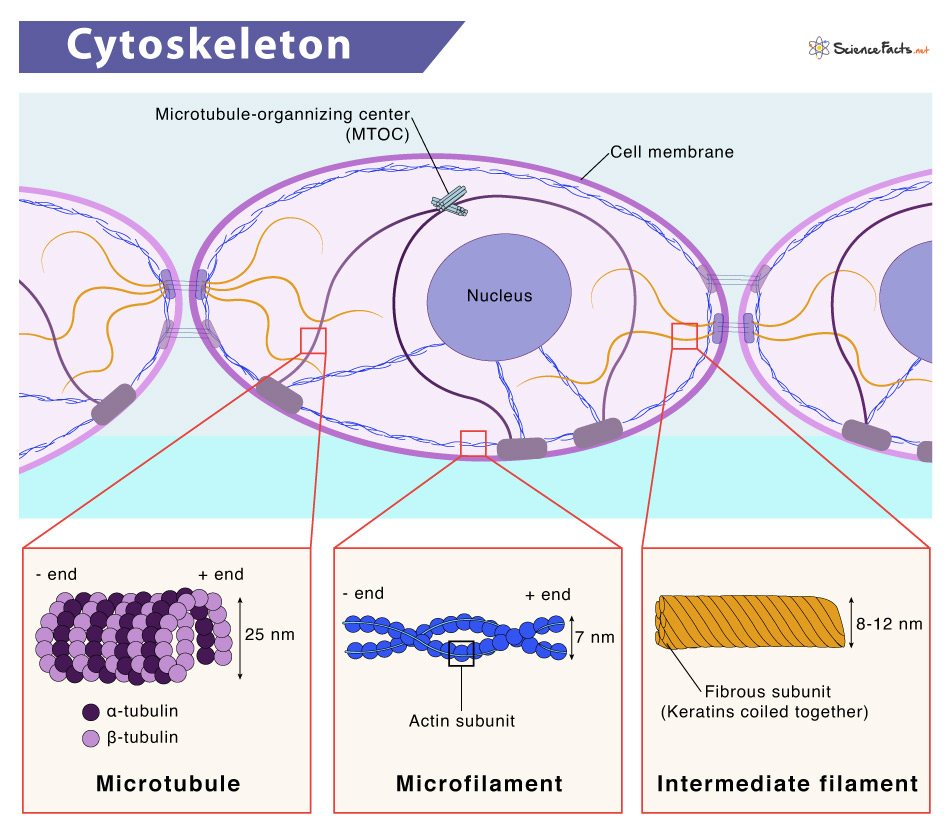
Just like the skeletal system of our body, the cytoskeleton is the skeletal system of a cell. Like our skeletal system, it provides a structural framework to the cell, helping it move and carry out several other activities.
Where is the Cytoskeleton Located
Parts and Structure
Functions: What does the Cytoskeleton Do
Microfilaments
Of the three types of cytoskeletal protein fibers, microfilaments are the narrowest, with a diameter of about 7 nm. They are made up of several intertwined actin monomers, resembling a double helix. As actin monomers are the principal component of microfilaments, they are also known as actin filaments. Like actins, microfilaments also have directionality, i.e., structurally different plus and minus end. Microfilaments provide mechanical support to the cell by forming a band just beneath the cell membrane. These filaments play several vital roles in the cell.
This network of filaments, linked to the plasma membrane by particular connector proteins, provides the shape and structure to the cell.They participate in specific cell junctions by linking transmembrane proteins to cytoplasmic proteins.They act as routes for the movement of a motor protein called myosin. Due to its relationship to myosin, it gets involved in many cellular events that require motion.They anchor the centrosomes at opposite poles of the cell during mitosis.During cytokinesis, in animal cell division, a ring composed of actin and myosin pinches the cell apart, producing two new daughter cells.In muscle cells, actin and myosin overlap each other, forming sarcomeres. When these filaments of a sarcomere slide against each other in concert, muscles contract. Hence, actinin forms the contractile part of the cytoskeleton.Microfilaments also serve as highways inside the cell to transport several cargoes, including protein-containing vesicles and other organelles. Different myosin motors help in shipping these cargoes by walking along the actin filaments.These filaments play an essential role in cell motility through lamellipodia, filopodia, or pseudopodia. For example, the crawling of a white blood cell (diapedesis) to reach the injured site.
Intermediate Filaments
Intermediate filaments (IFs) are a kind of cytoskeletal element comprised of dimers or two anti-parallel helices of varying protein sub-units, such as vimentin, glial fibrillary acidic protein, neurofilament proteins, keratins, and nuclear lamins. All the proteins are found in the cytoplasm except lamins, which are present in the nucleus, supporting the nuclear envelope. As the name suggests, their average diameter lies between microfilaments and microtubules, ranging from 8 to 12 nm. Some of IFs functions are:
To play an essential role in providing structural stability to the cellTo maintain the cell’s shape and anchor the nucleus and other organelles in place since they can bear the tension
Microtubules
Microtubules are the largest cytoskeletal component. These polymers, made up of globular protein subunits called α-tubulin and β-tubulin, occur throughout the cytoplasm. Microtubules are not only found in eukaryotic cells but some bacteria as well. As the name suggests, they are tiny hollow tubes with diameters ranging from 200 nm to 25 micrometers. Like microfilaments, they also exhibit polarity. In animal cells, microtubules originate from the microtubule-organizing center (MTOC), i.e., centrioles and basal bodies. They are the structural elements of flagella, cilia, and centrioles. There are three subgroups of microtubules: A few of their essential functions are listed below:
Their functions are associated with providing intracellular shape, locomotion, and transport.They give rise to the spindle apparatus, separating sister chromatids for equal distribution to each daughter cell during cell division.They provide the cell shape, thus resist compression.They help in intracellular transportation by providing a track along which vesicles move through the cell.They aid in the formation of the cell wall in plant cells.
Maintain the shape and provide support to the cellHolds a variety of cellular organelles in placeAssists in vacuole formationEnables internal and overall cell mobility. For instance, transportation of vesicles in and out of a cell, chromosomal movement during meiosis and mitosis, and migration of organelleCommunicates signals between cellsForms cellular protrusions such as cilia and flagella in some cells
